Taxonomy and phylogeny of prokaryotes present problems because of extensive horizontal gene transfer.
Horizontal gene transfer (HGT) or lateral gene transfer (LGT) is the movement of genetic material between unicellular and/or multicellular organisms other than by the ("vertical") transmission of DNA from parent to offspring. HGT is an important factor in the evolution of many organisms.
Bites from the insect Reduviidae (assassin bug) can, via a parasite, infect humans with the trypanosomal Chagas disease, which can insert its DNA into the human genome. It has been suggested that lateral gene transfer to humans from bacteria may play a role in cancer.
horizontal gene transfer in humans
Horizontal gene transfer and the origin of species: … - de la Cruz - Cited by 510
Evidence for Extensive Resistance Gene Transfer … - Shoemaker - Cited by 458
Horizontal gene transfer: building the web of life - Soucy - Cited by 221
Image result for horizontal gene transfer in humans Bites from the insect Reduviidae (assassin bug) can, via a parasite, infect humans with the trypanosomal Chagas disease, which can insert its DNA into the human genome. It has been suggested that lateral gene transfer to humans from bacteria may play a role in cancer. Horizontal gene transfer - Wikipedia https://en.wikipedia.org/wiki/Horizontal_gene_transfer Feedback About this result People also ask How does horizontal gene transfer occur? Horizontal gene transfer may occur via three main mechanisms: transformation, transduction or conjugation. Transformation involves uptake of short fragments of naked DNA by naturally transformable bacteria. Transduction involves transfer of DNA from one bacterium into another via bacteriophages. Horizontal Gene Transfer — Antimicrobial Resistance Learning Site ... https://amrls.cvm.msu.edu/.../acquisition-of-antimicrobial-resistance-via-horizontal-gene... Search for: How does horizontal gene transfer occur? Can horizontal gene transfer occur in humans? Bacteria and Humans Have Been Swapping DNA for Millennia. ... This DNA-sharing process, known as horizontal or lateral gene transfer (LGT), is now understood to occur by the direct movement of DNA between two organisms.Oct 1, 2016 Lateral Gene Transfer Between Humans and Microbes | The Scientist ... https://www.the-scientist.com/?articles.view/articleNo/47125/title/...and-Humans... Search for: Can horizontal gene transfer occur in humans? Is horizontal gene transfer evolution? Although gene exchange is easier in closely related organisms, horizontal gene transfer occurred between both domains in the evolution of Archaea and Bacteria. However, it is a disputed point whether horizontal gene transfer precludes the reconstruction of phylogenetic relationships in the microbial world.Oct 28, 2009 Horizontal gene transfer in evolution: facts and challenges ... rspb.royalsocietypublishing.org/content/277/1683/819 Search for: Is horizontal gene transfer evolution? Is binary fission horizontal gene transfer? Vertical gene transfer refers to the transfer of genetic material from a parent to the offspring. Vertical gene transfer only occurs through reproduction. Bacteria divide asexually through binary fission , which is a simple three step process: The bacterial chromosome replicates. Bacterial gene transfer - ToKToL https://www.toktol.com/notes/section/1163/biology/bacteria/bacterial-gene-transfer Search for: Is binary fission horizontal gene transfer? How does horizontal gene transfer work? Horizontal gene transfer is made possible in large part by the existence of mobile genetic elements, such as plasmids (extrachromosomal genetic material), transposons (“jumping genes”), and bacteria-infecting viruses (bacteriophages). ... In transduction, DNA is transmitted from one cell to another via a bacteriophage. Horizontal gene transfer | genetics | Britannica.com https://www.britannica.com/science/horizontal-gene-transfer Search for: How does horizontal gene transfer work? How is horizontal gene transfer different from vertical gene transfer? What is a lateral gene transfer? How does horizontal gene transfer contribute to antibiotic resistance? What is gene transfer? Who discovered horizontal gene transfer? How can genes be transferred between bacteria? How does horizontal gene transfer complicate phylogenetic trees? How genes are transferred from one organism to another? What are the two ways that bacteria can acquire antibiotic resistance?
When yeast cells are grown in liquid cultures, they metabolize glucose predominantly by glycolysis, releasing ethanol in the medium. When glucose becomes limiting, the cells enter diauxic shift characterized by decreased growth rate and by switching metabolism from glycolysis to aerobic utilization of ethanol.
The lac operon (lactose operon) is an operon required for the transport and metabolism of lactose in Escherichia coli and many other enteric bacteria. Although glucose is the preferred carbon source for most bacteria, the lac operon allows for the effective digestion of lactose when glucose is not available through the activity of beta-galactosidase.[1] Gene regulation of the lac operon was the first genetic regulatory mechanism to be understood clearly, so it has become a foremost example of prokaryoticgene regulation. It is often discussed in introductory molecular and cellular biology classes for this reason. This lactose metabolism system was used by Jacob and Monod to determine how a cell knows which enzyme to synthesize. Their work on the lac operon won them the Nobel Prize in Physiology in 1965 attempts to integrate our data and understanding, an area known as systems biology. Whereas classical biochemistry made great contributions to demonstrating the properties of proteins in isolation, our job now is to put things back together. Chapter 11, Systems Biology, presents a description of biological organization that is based on networks. Cells contain parallel sets of networks based on physical and logical interactions among molecules. Each network also has static and dynamic aspects. The ultimate, most profound, goal is a complete and integrated picture of all of life’s activity, from the molecule to the biosphere.
Palaeogenomics
English
Etymology
palaeo- + genomics
Noun
palaeogenomics
The study of ancient genomes, especially those of extinct organisms
In many ways, palaeogenomics began when the first ancient DNA sequence was reported. This first sequence was derived from a stuffed museum specimen of the quagga, an extinct mammal related to the zebra. Unspecified and unselected DNA was extracted from the quagga specimen, cloned into a bacterial library, and then sequenced. It took another 17 years and the development of PCR before two independent groups successfully sequenced the complete mitochondrial genomes from several extinct moa species. Only 4 years later, using the original approach of cloning nonspecific ancient DNA extract and shotgun sequencing, the first ancient nuclear DNA sequences were determined, this time from the extinct cave bear. Since these early successes, palaeogenomics has rapidly expanded, because of both technological development and increasing interest in ancient DNA research
the important ideas and tools
– of taxonomy and phylogeny, on the classical species
level and on the molecular level
"There is nothing boring in the human genome kindergartners reading this information. we're just —ERIC LANDER ACGT Acquired Immunodeficiency Sy Adenine Allele Amino Acids Ancestry-informative Markers Animal Model Antibody Anticodon Antisense Apoptosis Autism Autosomal Dominant
Although clinical applications are undoubtedly the most important, genomics has important contributions to make to
human palaeontology,
anthropology,
the law.
Cancer genomics – the comparison of sequences from normal and tumour cells from single patients – has become a major activity.
Abnormal splicing variants are also thought to contribute to the development of cancer, and splicing factor genes are frequently mutated in different types of cancer. synteny blocks
In classical genetics, synteny describes the physical co-localization of genetic loci on the same chromosome within an individual or species. Today, however, biologists usually refer to synteny as the conservation of blocks of order within two sets of chromosomes that are being compared with each other.
Metagenomics is the study of genetic material recovered directly from environmental samples. The broad field may also be referred to as environmental genomics, ecogenomics or community genomics. While traditional microbiology and microbial genome sequencing and genomics rely upon cultivated clonalcultures, early environmental gene sequencing cloned specific genes (often the 16S rRNA gene) to produce a profile of diversity in a natural sample. Such work revealed that the vast majority of microbial biodiversity had been missed by cultivation-based methods.[1] Recent studies use either "shotgun" or PCR directed sequencing to get largely unbiased samples of all genes from all the members of the sampled communities.[2] Because of its ability to reveal the previously hidden diversity of microscopic life, metagenomics offers a powerful lens for viewing the microbial world that has the potential to revolutionize understanding of the entire living world.[3] As the price of DNA sequencing continues to fall, metagenomics now allows microbial ecology to be investigated at a much greater scale and detail than before.
The "surgeon left an instrument / or a gauze, swab or his wristwatch inside a patient" during the operation thoseHeadlines such as this can be devastating and can really break a surgeons career. Many years of hard work you put in to become a good surgeon is lost due to a few minutes of the carelessness of the surgical team.
Surgeons call them 'nevers' because they're mistakes they should NEVER make: like leaving things inside of you during surgery. But they do happen.. all the time!
Although it is a systems fault and the onus is on the whole surgical team the surgeon carries the vicarious responsibility as he is supposedly the leader of the team
I have worked both as a general surgeon and a pediatric surgeon in India from 1982 to 1994
after which came to the USA and switched careers and became an Internist.
We are all aware that peoples personality determines their behavior.
Behavior changes peoples personality
Peoples personality determines their behavior.
Before coming to the USA when I wwas asurgeon in India. I used to be always hyperactive
abrupt having little patience for anyone and was supremely confident that what I say and do was the best thing for the patient
Known in medical terminology as the unintended retention of foreign objects (URFOs) or retained surgical items (RSIs), this is a serious patient safety issue that can cause death or harm patients physically and emotionally.
We in India do not have a registry or a reporting system so we have no statistics except the sensational news reports or the occasional Consumer forum /court decisions in such cases.
The Joint Commission has received more than 770 voluntary reports of URFOs in the past seven years. These cases resulted in 16 deaths, and about 95 percent of these incidents resulted in additional care and/or an extended hospital stay. Beyond the human toll, studies have shown that objects left behind after surgery may cost as much as $200,000 per case in medical and liability payments.”.
\
The following are just a few of the recommended actions outlined by the Joint Commission:
· Create a highly reliable and standardized counting system to prevent URFOs – making sure all surgical items are identified and accounted for.
· Develop and implement effective evidence-based organization-wide standardized policy and procedures for the prevention of URFOs through a collaborative process promoting consistency in practice to achieve zero defects. ·
Appropriate documentation should include the results of counts of surgical items, instruments, or items intentionally left inside a patient (such as needle or device fragments deemed safer to remain than remove), and actions are taken if count discrepancies occur.
Tracking discrepant counts is important to understanding practical problems. the joint commision says it was 770 patients over a 7 year period where as this sensational reporting, where a picture with a large tailor'sscissors super imosed on a chest xray, 9 couldnt find a surgical instrument !) says it is 1500 every year !
For more: http://bit.ly/1c7fYv7
According to the sentinel event data, the most common root causes of URFOs reported to The Joint Commission are:
• The absence of policies and procedures • Failure to comply with existing policies and procedures • Problems with hierarchy and intimidation ( There used to be two CTVS surgeons in AIIMS who were famous for making residents and fellows literally shiver and piss in their pants) • Failure in communication with physicians • Failure of staff to communicate relevant patient information • Inadequate or incomplete education of staff
In India especially in Hyderabad, it is " chalta hai " attitude about everything
www.jointcommission.org Published for Joint Commission accredited organizations and interested health care professionals, Sentinel Event Alert identifies specific types of sentinel and adverse events and high risk conditions, describes their common underlying causes, and recommends steps to reduce risk and prevent future occurrences. Accredited organizations should consider information in an Alert when designing or redesigning processes and consider implementing relevant suggestions contained in the Alert or reasonable alternatives. Please route this issue to appropriate staff within your organization. Sentinel Event Alert may only be reproduced in its entirety and credited to The Joint Commission. To receive by email, or to view past issues, visit www.jointcommission.org. __________________________ A complimentary publication of Issue 51, October 17, 2013 The Joint Commission Preventing unintended retained foreign objects The unintended retention of foreign objects (URFOs) – also called retained surgical items (RSIs) – after invasive procedures can cause death, and surviving patients may sustain both physical and emotional harm, depending on the type of object retained and the length of time it is retained. There may be an extended time frame between occurrence and detection of an URFO. Retained foreign objects are most commonly detected immediately post-procedure; by X-ray; during routine follow-up visits; or from the patient’s report of pain or discomfort. URFOs refer to any item or foreign object related to any operative or invasive procedure that is left inside a patient.1 Objects most commonly left behind after a procedure are: • Soft goods, such as sponges and towels • Small miscellaneous items, including unretrieved device components or fragments (such as broken parts of instruments), stapler components, parts of laparoscopic trocars, guidewires, catheters, and pieces of drains • Needles and other sharps • Instruments, most commonly malleable retractors1 A New York Times article published in September 2012 illustrates the adverse effects of an URFO. Four years after having a hysterectomy, a woman in Kentucky began to experience severe abdominal pain. A CT scan revealed a surgical sponge left behind by the surgical team that had performed the hysterectomy. Upon surgical exploration, the retained sponge was found to have caused a serious infection, which required bowel resection. The patient suffered from severe health issues, anxiety, depression, disability and social isolation.2 Not only does an URFO harm the patient, it adds significantly to the average total cost of caring for the patient. In a recent review, the Pennsylvania Patient Safety Authority estimated that the average total cost of care related to an URFO is about $166,000.3 This cost includes legal defense, indemnity payments, and surgical costs not reimbursed by the Centers for Medicare & Medicaid Services. Another study estimated medical and liability costs to be $200,000 or more per incident.4 Events, risk factors and root causes From 2005 to 2012, 772 incidents of URFOs were reported to The Joint Commission’s Sentinel Event database.* Sixteen deaths resulted from these incidents. About 95 percent of these incidents resulted in additional care and/or an extended hospital stay. In hospital settings, these incidents occurred in operating rooms, labor and delivery areas, as well as ambulatory surgery centers and other areas where invasive procedures are performed (e.g., cath lab, GI lab, interventional radiology, emergency department). According to the sentinel event data, the most common root causes of URFOs reported to The Joint Commission are: • The absence of policies and procedures • Failure to comply with existing policies and procedures * The reporting of most sentinel events to The Joint Commission is voluntary and represents only a small proportion of actual events. Therefore, these data are not an epidemiologic data set and no conclusions should be drawn about the actual relative frequency of events or trends in events over time. Sentinel Event Alert, Issue 51 Page 2 www.jointcommission.org • Problems with hierarchy and intimidation • Failure in communication with physicians • Failure of staff to communicate relevant patient information • Inadequate or incomplete education of staff According to one study, the most common risk factors for URFOs include: patients with high body mass index (risk ratio for each one-unit increment, 1.1 [95 percent confidence interval, 1.0 to 1.2]); an emergent or urgent procedure; and unanticipated/ unexpected change during the procedure.5 (Some examples of changes that can occur during a procedure include: a change in approach/incision, type of procedure, added procedure, or the development of a complication during the procedure). Other risk factors include intraabdominal surgery; more than one surgical procedure; involvement of multiple surgical teams; multiple staff turnovers during the procedure; and unexpected intraoperative development. Occurrence of an URFO was nine times as likely when an operation was performed on an emergency basis and four times as likely when the procedure changed unexpectedly (see examples of change above). 3 An additional risk factor is long procedure duration.6,7 URFOs also occur in patients who exhibit none of these risk factors. In order to prevent retained surgical items and sponges, surgeons and operating room staff have traditionally relied on “cavity sweeps” and manual counting protocols – both of which are prone to human error. Current practices for counting sponges have a 10 to 15 percent error rate.8 In addition, 80 percent of retained sponges occur with what staff believe is a correct count. 8 Sentinel event data show an incorrect or “discrepant” count in 52 of the 772 URFO sentinel events reported to The Joint Commission. The Pennsylvania Patient Safety Authority’s Reporting System database shows 22.3 percent of URFOs were associated with a discrepant count.9 Many counting procedures lack the elements of high reliability but are entrenched and difficult to change, said Verna C. Gibbs, M.D., professor of clinical surgery, University of California, San Francisco, and director of No Thing Left Behind® , a national surgical patient safety project to prevent retained surgical items. High reliability science studies organizations such as those in the commercial aviation industry, which manage great hazard extremely well, and in which the goal is zero harm. In order to achieve high reliability, leadership must commit to this goal; the culture must support workers who identify and report unsafe conditions; and systematic quality improvement approaches need to be implemented that reliably measure the magnitude of the problem (e.g., days between procedures with an URFO), identify the contributing factors and root causes, and develop solutions for the most important causes.10,11 Studies show that the risk of URFOs is significantly reduced following improvements to counting procedures. Team members need to move from varying practices to standardized practices – to develop and sustain reliable counting practices that ensure all surgical items are accounted for (i.e., are reconciled).12 One children’s hospital reduced the number of reported incorrect counts and count discrepancies by 50 percent between 2009 and 2010, and also improved its entire count process.13 Recommendations and potential strategies for improvement Guidelines, processes and tools have become available to help team members develop riskreduction strategies that can be adopted and followed organization-wide.1,12,14 These strategies include improved multi-stakeholder perioperative processes, enhanced team communication, and the use of assistive technology.1,12,14,15,16 Organizations should provide continuous education or training to appropriate staff about new and existing policies and procedures that are in place to prevent URFOs. The following recommendations and potential strategies can be used to help prevent URFOs. Should your organization discover and remove an URFO, follow your organization’s established policy for reporting, analyzing and communicating the event to staff and the patient and his or her family. Effective processes and procedures 1. Create a highly reliable and standardized counting system to prevent URFOs – making sure all surgical items are identified and accounted for. The counting system should be supported by organizational leaders, and developed using a multidisciplinary approach, involving surgeons, proceduralists, nurses, surgical technologists, anesthesiologists, radiologists, and radiology technologists working together as a team in an environment that promotes the exchange of knowledge and information.1,12,14,16,17 2. Develop and implement effective evidencebased organization-wide standardized policy and procedures for the prevention of URFOs through a collaborative process promoting consistency in practice to achieve zero defects. Use resources published by The Joint Commission,17 World Health Organization, American College of Surgeons,14 Association of periOperative Sentinel Event Alert, Issue 51 Page 3 www.jointcommission.org Registered Nurses,12 No Thing Left Behind, and other organizations and publications as a guide. The policy should apply to all operative and other invasive procedures, and should address the following. A counting procedure should: • Be performed audibly and visibly by two persons engaged in the process, usually scrub tech or scrub nurse and circulating registered nurse. The surgical team should verbally acknowledge verification of the count. • Include counts of items added to the surgical field throughout the surgery or procedure. • Include counts of soft goods (including therapeutic packing), needles/sharps, instruments, and small miscellaneous items, and document unretrieved device fragments.1,12 • Verify that counts printed on prepackaged sponges and instrument sets are correct.1,12 Handle the discrepancy per the organization’s policy. • Be performed before the procedure begins, in order to establish a baseline count; before the closure of a cavity within a cavity; before wound closure begins; at skin closure or end of procedure; and at the time of permanent relief of either the scrub person or the circulating registered nurse.18 • Be applicable in all settings where invasive procedures are performed. • Be reviewed periodically and revised as appropriate.12,14 Wound opening and closing procedures should include: • Inspection of instruments for signs of breakage – before and after use – to prevent the retention of device fragments.19 • Adherence to the organization’s established counting procedure. • Methodical wound exploration,1,14 including visual and, whenever possible, manual examination.20 This can and should be done for laparoscopic procedures as well. • Empowerment of any member of the operative team to call a “closing time out” prior to the initial closing count to allow for an uninterrupted count. Intra-operative radiographs should be performed: • When the surgical count is “incorrect” (i.e., discrepant).The entire surgical field should be radiographed, and it should be interpreted by a physician at the completion of the operative procedure, prior to the patient’s transfer from the OR. Ensure direct communication between the surgical team and radiologist. The requisition should include the name of the missing item and the results of the radiologic image should be directly communicated to the surgical team. • When the operative procedure is determined by the surgical team to be at high risk for retained surgical items, even though methodical wound exploration has been performed and the surgical item count is correct. • If counts remain unreconciled after initial radiologic examination, the surgical team should consider additional imaging or further wound exploration.12,21 Effective communication 3. Institute team briefings and debriefings as a standard part of the surgical procedure to allow the opportunity for any team member to express concerns they have regarding the safety of the patient, including the potential for an URFO. This will promote open communication among surgical team members. Examples: Before the procedure or as part of the time out, the surgeon could remind the team that the patient or procedure is at risk for an URFO; during the procedure, a white board could be used to display the count and to help foster team awareness and shared responsibility; 22,23 at the end of the procedure, team members can raise or be asked about any concerns related to the procedure or the patient’s recovery.21 Team training, based on crew resource management (CRM), is effective in promoting assertiveness and overcoming hierarchical barriers to communication. 4. Ensure that the surgeon verbally verifies the results of the counting procedure. Appropriate documentation 5. Document the results of counts of surgical items, instruments, or items intentionally left inside a patient (such as needle or device fragments deemed safer to remain than remove), and actions taken if count discrepancies occur.14 Tracking discrepant counts is important to understanding practical problems; tracking reports and data also can be discussed at improvement meetings. Collecting, analyzing and sharing accurate data is key to understanding your organization’s frequency or risk of URFOs, identifying the types of URFOs that occur most frequently, and determining how to address certain kinds of URFOs. Safe technology 6. Research the potential of using assistive technologies1,12,14,16,24,25 to supplement manual counting procedures and methodical wound exploration. More commonly used technologies Sentinel Event Alert, Issue 51 Page 4 www.jointcommission.org include bar-coding to aid counting, radio-opaque material or radiofrequency (RF) tags to detect technology-enabled soft goods, and radio frequency identification (RFID) systems to aid counting and detection.8,26 Related Joint Commission requirements The unintended retention of a foreign object in a patient after surgery or other invasive procedure is considered a reviewable sentinel event by The Joint Commission. Accredited organizations are expected to respond to sentinel events as part of a patient safety program outlined in the following standards and elements of performance (EP) for hospitals, ambulatory and office-based surgery facilities. LD.04.01.07: The organization has policies and procedures that guide and support patient care, treatment and services. LD.04.04.05: The organization has an organization-wide, integrated patient safety program [within its performance improvement activities]. (Wording in brackets is added for hospitals only.) EP 5: As part of the safety program, the leaders create procedures for responding to system or process failures. EP 7: The leaders define “sentinel event” and communicate this definition throughout the organization. EP 8: The organization conducts thorough and credible root cause analyses in response to sentinel events as described in the "Sentinel Events" (SE) chapter of the manual. EP 9: The leaders make support systems available for staff who have been involved in an adverse or sentinel event. RI.01.02.01: The organization respects the patient’s right to participate in decisions about his or her care, treatment, and services. EP 21: The organization informs the patient or surrogate decision-maker about unanticipated outcomes of care, treatment, and services that relate to sentinel events considered reviewable by The Joint Commission. EP 22: [Hospitals only] The licensed independent practitioner responsible for managing the patient's care, treatment, and services, or his or her designee, informs the patient about unanticipated outcomes of care, treatment, and services related to sentinel events when the patient is not already aware of the occurrence or when further discussion is needed. Resources WHO guidelines for safe surgery 2009: safe surgery saves lives: See Objective 7: The team will prevent inadvertent retention of instruments and sponges in surgical wounds, and Objective 9: The team will effectively communicate and exchange critical information for the safe conduct of the operation World Health Organization: Surgical Safety Checklist Pennsylvania Patient Safety Authority: Retained Foreign Object Audit Form References 1 NoThing Left Behind®: A national surgical patient-safety project to prevent retained surgical items. www.nothingleftbehind.org (accessed March 18, 2013) 2 O’Connor A: When surgeons leave objects behind. The New York Times, September 24, 2012 3 Beyond the count: preventing retention of foreign objects. Pennsylvania Patient Safety Advisory, June 2009;6(2):39-45, http://patientsafetyauthority.org/ADVISORIES/AdvisoryLi brary/2009/Jun6(2)/Pages/39.aspx (accessed May 16, 2013 4 Regenbogen, et al: Prevention of retained surgical sponges: A decision-analytic model predicting relative cost-effectiveness. Surgery, 2009;145:527-35 5 Gawande AA, et al: Risk factors for retained instruments and sponges after surgery. New England Journal of Medicine, January 16, 2003;348(3)229-35 6 Stawicki S, et al: Retained surgical items: a problem yet to be solved. Journal of the American College of Surgeons, January 2013;216(1):15-22 7 Judson, TJ, et al: Miscount incidents: A novel approach to exploring risk factors for unintentionally retained surgical items. The Joint Commission Journal on Quality and Patient Safety, October 2013;39(10):468-473 8 Tips for reducing retained surgical items. Same-Day Surgery, September 2009;91-92 9 Martindell D: Update on the prevention of retained surgical items. Pennyslvania Patient Safety Advisory, September 2012;9(3):106-110, http://patientsafetyauthority.org/ADVISORIES/AdvisoryLi brary/2012/Sep;9(3)/Pages/106.aspx (accessed May 16, 2013 10 Chassin M, Loeb J: The ongoing quality improvement journey: Next stop, high reliability. Health Affairs, April 2011;30(4), http://content.healthaffairs.org/content/30/4/559.full?ijkey =UoA7j1SNli6pQ&keytype=ref&siteid=healthaff (accessed May 14, 2013) 11 Chassin MR, Loeb JM: High-reliability health care: Getting there from here. The Milbank Quarterly, September 2013;91(3):459-490, http://www.jointcommission.org/highreliability_health_care_getting_there_from_here/ (accessed September 25, 2013) 12Goldberg JL, Feldman, DL: Implementing AORN recommended practices for prevention of retained surgical items. AORN Journal, February 2012;95(2):205- 219 Sentinel Event Alert, Issue 51 Page 5 www.jointcommission.org 13 Norton EK, Micheli, AJ: Patients count on it: An initiative to reduce incorrect counts and prevent retained surgical items. AORN Journal, January 2012;95(1) 14 American College of Surgeons: Statement on the prevention of retained foreign bodies after surgery. 2005;90(10):15 15 Cima RR, et al: Incidence and characteristics of potential and actual retained foreign object events in surgical patients. Journal of the American College of Surgeons, 2008;207:80-87 16 Cima RR, et al: A multidisciplinary team approach to retained foreign objects. The Joint Commission Journal on Quality and Patient Safety, March 2009;35(3):123-132 17 The Joint Commission: Resources for managing hospital-acquired conditions. Foreign objects retained after surgery, http://www.jcrinc.com/foreign-objectsretained-after-surgery (accessed March 18, 2013) 18 Department of Veterans Affairs, Veterans Health Administration: VHA Directive 2010-017. Washington, DC, http://www.va.gov/vhapublications/ViewPublication.asp?p ub_ID=2186 (accessed June 20, 2013) 19 U.S. Food and Drug Administration, Public health notification; unretrieved device fragments. January 15, 2008, http://www.fda.gov/MedicalDevices/Safety/AlertsandNotic es/PublicHealthNotifications/ucm062015.htm (accessed April 15, 2013) 20 Gibbs VC: Sponge ACCOUNTing System. Methodical Wound Examination. Online supplement to Same-Day Surgery, September 2009 21 World Health Organization: WHO guidelines for safe surgery 2009: safe surgery saves lives. Geneva, Switzerland, 2009;pp. 72-75, 78-82, http://whqlibdoc.who.int/publications/2009/978924159855 2_eng.pdf (accessed May 24, 2013) 22 ECRI Institute: The case of the missing sponge: Practice variation is culprit. Risk Management Reporter, June 2012 23 Edel, E: Increasing patient safety and surgical team communication by using a count/time out board. AORN Journal, October 2010;92:4 24 White SV, interviewer: Interview with a quality leader: Dr. Verna Gibbs on surgical safety. Journal for Healthcare Quality, November/December 2012;34(6):21- 26 25 Asiyanbola, et al: Prevention and diagnosis of retained foreign bodies through the years: past, present and future technologies. Technology and Health Care, 2012;20(5):379-386 26 Steelman, VM and Cullen, JJ: Designing a safer process to prevent retained surgical sponges: A healthcare failure mode and effect analysis. AORN Journal, August 2011;94(2):132-141 _________________________________________________ Patient Safety Advisory Group The Patient Safety Advisory Group informs The Joint Commission on patient safety issues and, with other sources, advises on topics and content for Sentinel Event Alert. Members: James P. Bagian, M.D., P.E. (chair); Michael Cohen, R.Ph., M.S., Sc.D. (vice chair); Paul W. Abramowitz, Pharm.D., FASHP; Jane H. Barnsteiner, R.N., Ph.D., FAAN; James B. Battles, Ph.D.; William H. Beeson, M.D.; Patrick J. Brennan, M.D.; Cindy Dougherty, R.N., B.S., CPHQ; Frank Federico, B.S., R.Ph.; Marilyn Flack; Steven S. Fountain, M.D.; Suzanne Graham, R.N., Ph.D.; Martin J. Hatlie, Esq.; Robin R. Hemphill, M.D., M.Ph.; Jennifer Jackson, B.S.N., J.D.; Paul Kelley, CBET; Heidi B. King, FACHE, BCC, CMC, CPPS; Jane McCaffrey, M.H.S.A., DFASHRM; Mark W. Milner, R.N., M.B.A., CPHQ, FACHE; Jeanine Arden Ornt, J.D.; Grena Porto, R.N., M.S., ARM, CPHRM; Matthew Scanlon, M.D.; Ronni P. Solomon, J.D.; Dana Swenson, P.E., M.B.A.
‘There are 600 potentially endangered languages in India… each dead language takes away a culture system’
In the interview, Ganesh Devy spoke about the dying and dead languages of India, how some languages gain popularity while others remain marginalised, and the impact of colonisation on the language system of India.
SHARE
Written by Adrija Roychowdhury | New Delhi | Updated: February 22, 2018 10:19:18 am
Click on the image for a larger view of the language tree
When renowned literary critic and activist Ganesh Narayan Devy set out to map the linguistic diversity of India, he had no inkling he would encounter languages that are barely known in the states in which they are spoken. Among his interesting discoveries were — 200 words describing snow in the Himalayan region alone, an old form of Portuguese spoken in villages close to Mumbai, a form of Japanese spoken in parts of Gujarat, and a language from Myanmar that is popular in the islands of Andaman.
Devy, who documented 780 Indian languages while conducting the People’s Linguistic Survey of India in 2010, also, shockingly, found that 600 of these languages were dying. He added close to 250 languages in India had already died over the past 60 years.
Ganesh Devy
When a language dies, as Devy notes, “a unique way of looking at the world disappears”. On the occasion of International Mother Language Day, the critic spoke to Indianexpress.com about the dying and the dead languages of India. In the interview, he also dwells on how some languages gain popularity while others remain marginalised, and the impact of colonisation on the language system of India.
Excerpts:
What are some of the dying and dead languages of India?
According to UNESCO, any language that is spoken by less than 10,000 people is potentially endangered. In India, after the 1971 census, the government decided that any language spoken by less than 10,000 people need not be included in the official list of languages. In India, therefore, all the languages that are spoken by less than 10,000 people are treated by the state as not worthy of mention and treated by the UNESCO as potentially endangered. As per my survey, there are close to 780 languages in India, out of which about 600 are potentially endangered. The census of 1991 and 2001 show not more than 122 languages. So, most others have to be called potentially endangered.
Examples of such languages would be Wadari, Kolhati, Golla, Gisari. These are languages of nomadic people in Maharashtra, Karnataka and Telangana. Then there several tribal languages as well, such as Pauri, Korku, Haldi, Mavchi. In Assam, there is Moran, Tangsa, Aiton. There seems to be about 250 languages that disappeared in the last 60 years. There used to be languages called Adhuni, Dichi, Ghallu, Helgo, Katagi. The Bo language in Andaman disappeared in 2010 and the Majhi language in Sikkim disappeared in 2015. But we need to remember that it is impossible to show a language dying in the last moment of its life. A language is not a single life system. It is a very large symbolic system. When the symbols collapse they do not do so in a single moment. The collapse is sprayed over a large time.
What happens when a language dies?
When a language dies, its speakers decide to migrate. First, they migrate to another language and then they physically start migrating to another region. The second thing that happens is that their traditional livelihood patterns go down. They may have some special skills and that disappears. Thirdly, a unique way of looking at the world disappears. Every language is a unique worldview.
How do we conserve a dying language?
It is very simple. We need to create livelihood support for the speakers of the language. If they have livelihood available within their language, nobody would want to switch from their language to any other language.
How do some languages gain popularity while others get marginalised?
There are a few major reasons for this. One is that some languages as against other alternate languages in the area gain popularity because of an easier syntax. For instance, in Hindi, you can say ‘ladka chalta hain’ and you can also say ‘chalta hain ladka.’ So the syntax is flexible. But it’s not always that easy in English. This is one major reason but not the reason always. Secondly, the social dominance by any group leads to the language of that group becoming more popular in that society. For instance, Sanskrit became popular in ancient India because of social domination by speakers of Sanskrit. Or English has become popular because of the colonial rule. Thirdly, when a language becomes useful in a marketplace, that language gains greater currency. For instance, we in India speak one language at home, maybe another in office, but when we go to the market we might use neither. For example in Delhi, you might use Punjabi or Bengali at home, English in the office, but in the marketplace, one tends to use the Hindi language. So the patterns of political domination, use in the marketplace and ease of syntactic structuring are three reasons why some languages become more popular than other languages.
What is the impact of colonisation on Indian languages?
Quite surprisingly, in other continents, the colonial impact wiped out the native languages. In India that did not happen. Our languages survived. However, the colonial times brought us the print technology and only very few of our languages got printed. The one that got printed eventually got states to themselves since in India our states are designed in linguistic terms. The other languages did not get states for them, they did not get official recognition and therefore became secondary citizens in the language republic of India.
How is the language we speak related to our worldview?
In every manner without any exception, the language we learn or use is the absolute condition of our narrative of the world and the way we see the world. There is no escape from it. A given language only has a certain kind of ability to narrate the world and the consciousness can enter the world only to the extent that languages can allow it to enter the reality surrounding it. If a language has seven terms for distributing colours, then the speaker of the language will see the world only in those colours. But if there is another language which has more colour terms, then the distribution of the world is more multicoloured. For instance, in Marathi, there is a colour term called Kirmizi that cannot be translated into any English term at all. It is brownish, greenish, bluish, it’s almost like the colour combination we see in a firefly. It is impossible to replicate that perception in the English language. But in the English language, we have navy blue or sky blue and many other languages might not have the exact colour term that translates the same. This is how language allows or disallows us in interpreting the world.


 SHARE
SHARE


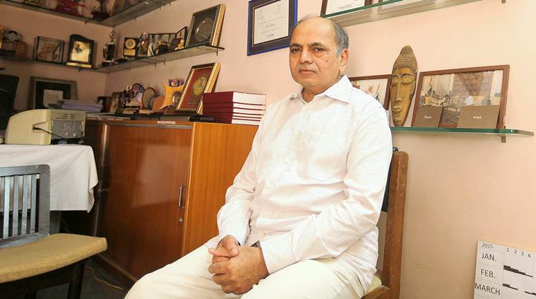 Ganesh Devy